Kind of infrastructure
Singular equipments
Typology
Imaging/microscopy
University
Pau et Pays de L'Adour (UPPA)
Hubs
Green Energies
Circular Economy and Environment
Global health
Application / Industrial sector
Energy & environment
Description of equipment
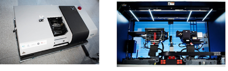
X ray tomography enables to visualise the three-dimensional microstructure of both opaque and transparent media at micrometer resolution in a non-invasive way. The DMEX Center for X-ray Imaging hosts four tomographs, respectively targeting high resolution, high contrast, high speed and large field-of-view applications. The center has particular experience in "in-situ" and "in-operando" studies, where the sample is imaged while being subjected to specific conditions (flow, mechanical loading, humidity, electric charge, etc.). One scanner is equipped with a spectral detector enabling non-invasive three-dimensional mapping of heavy elements.
Location
Service
UPPA Tech / DMEX
Features
Others tipologies
Materials / solid state / nuclear physics
Technicians
UPPA research engineers, Researchers
Equipment list
Bruker Skyscan, Zeiss XRadia 510, Tescan UniTOM XL Spectral, Tescan DynaTOM
Available measurements
Solide material morphology
Sample preparation
Plugs, on small pieces (10 cm size)
Deployment of the results
Analysis report, 3D model
Price list
On demand
Contact
Person name
Karine PINEL
Email
karine.pinel@univ-pau.fr
Phone
+33 (0)6 62 55 32 79